Glaucoma & Advanced Testing
At EyeTech Eye Center, we offer a wide array of cutting-edge diagnostic services to ensure the health and well-being of your vision. From visual field testing to detect central and peripheral dysfunction to advanced imaging techniques like fundus photography and optical coherence tomography (OCT), our technologies help identify and monitor a variety of eye conditions such as glaucoma, macular degeneration, and diabetic retinopathy. Additionally, specialized tests like corneal pachymetry and gonioscopy aid in evaluating corneal thickness and the angles within the eye for effective glaucoma diagnosis. Our team is dedicated to utilizing state-of-the-art methods, including Visual Evoked Potential Testing (VEP) and Electroretinography (ERG), to provide comprehensive assessments of both neurological and ocular health. Trust us to deliver precise and personalized eye care solutions tailored to your needs.
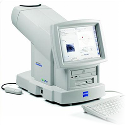
Visual Field Testing (VF)
A visual field test is an essential diagnostic procedure that evaluates the functionality of both central and peripheral vision. This test is instrumental in detecting abnormalities or dysfunctions, which could be indicative of glaucoma, neurological disorders, or other underlying health conditions affecting visual performance. By identifying these issues early, visual field testing enables healthcare professionals to take prompt and effective measures to preserve and improve vision, ensuring the overall well-being of the patient’s ocular health.
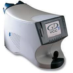
Glaucoma Diagnosis (GDx)
The GDx is a sophisticated diagnostic tool designed to scan the posterior of the eye, capturing detailed images of the optic nerve located at the back of the eye. By analyzing the thickness of the nerve fiber layer, the GDx provides critical insights into the health of the optic nerve. This advanced technology plays a vital role in the early detection of glaucoma, as it can identify signs of the disease long before individuals experience any noticeable symptoms of vision loss. Through its precise and proactive approach, the GDx enables timely diagnosis and intervention, helping to protect and preserve vision.
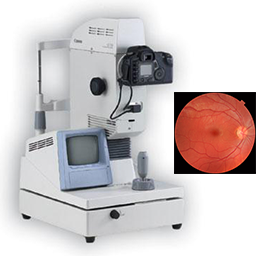
Fundus Photography – Retinal Imaging
Fundus Photography is the photography of the interior surface of the eye. The photograph provides us with an in-depth view of the retinal surface where many eye diseases first manifest. This photographic examination allows us to screen for ocular problems such as macular degeneration, glaucoma, retinal holes/detachments, and many symptoms of systemic diseases including diabetes and hypertension. The photograph is stored in your medical records for future comparisons to detect any changes over time.
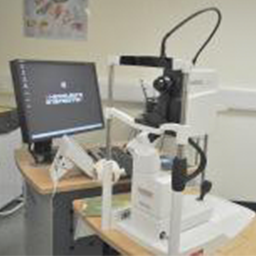
Optical Coherence Tomography (OCT)
Optical Coherence Tomography (OCT) is an advanced diagnostic tool that delivers high-resolution, cross-sectional imaging of the structures at the back of the eye. This cutting-edge technology allows for the detailed examination and detection of changes in the retina and other areas critical to vision. OCT is instrumental in identifying and monitoring various eye diseases, including glaucoma, diabetic retinopathy, and macular edema. By providing precise imaging, it enables eye care professionals to assess the progression of these conditions effectively and tailor treatment plans to safeguard and improve visual health.
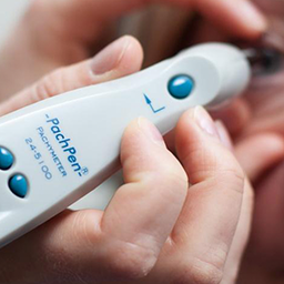
Corneal Pachymetry
Pachymetry is a specialized diagnostic procedure that utilizes a medical device called a Pachymeter to precisely measure the thickness of the cornea. This important test is commonly performed for individuals suspected of having glaucoma, as corneal thickness can influence eye pressure readings and aid in diagnosis. Additionally, pachymetry is an essential tool in screening for Keratoconus, a condition that affects the shape and integrity of the cornea, and is also crucial for those considering LASIK surgery, ensuring the cornea meets the necessary criteria for a safe and effective procedure.
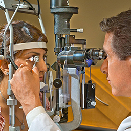
Gonioscopy
Gonioscopy is a specialized diagnostic procedure that employs a Goniolens and a slit lamp to measure the angle between the cornea and the iris within the eye. This precise evaluation is critical for identifying and diagnosing eye conditions linked to glaucoma, as it helps assess the drainage system of the eye and detect potential abnormalities. By providing valuable insight into the eye’s internal structures, gonioscopy plays a vital role in glaucoma management and treatment planning, ensuring optimal care for patients at risk.
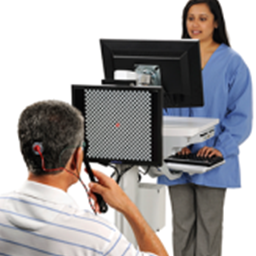
Visual Evoked Potential Testing (VEP) and Electroretinography (ERG)
Visual Evoked Potential measures the evoked potential or signals in the brain in response to a stimulus. It measures and records the amount of electrical activity and speed of the signals the brain, primarily the visual cortex, is sending and receiving. Similar to VEP, Electroretinography measures the electrical responses of the cells in the retina. Both the VEP and ERG tests provide comprehensive information for the diagnosis of eye diseases such as glaucoma, macular degeneration, and other neurological or eye conditions..
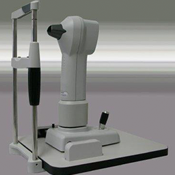
Corneal Topography and Imaging
Corneal Topography is an advanced imaging technique specifically designed to map the surface curvature of the cornea in intricate detail. This technology is crucial for identifying irregularities in the corneal shape, which can affect vision and overall eye health. Corneal Topography is commonly used to determine if a patient is an ideal candidate for Corneal Refractive Therapy (CRT) by providing essential data about the cornea’s structure. Additionally, it plays a key role in evaluating and optimizing the fit of CRT lenses and other contact lenses to ensure comfort and effectiveness, supporting the best possible visual outcomes for patients.
